Due to reduced intravascular volume( e.g overdiuresis, edematous states, diarrhoea,surreptitious vomitting) or Barter's syndrome where there's defect in kidneys' ability to conserve sodium.
There's:
high aldosterone
hypokalaemia
muscle weakness
metabolic alkalosis
high renin (renin dependent hyperaldosteronism)
Unlike 1° aldosteronism,
There's no hypertension
There may be edema
Saturday, May 5, 2012
Sensitivity and screening
A good screening test is very sensitive. This means it picks up most cases of the disease so a negative result tells you with good level of certainty that the disease is absent. Thus a good screening test "rules out". It therefore also has high number of false positives.
It is usually not very specific, i.e if it is positive, it may be due to something other than the disease but it must be very sensitive i.e if it is negative, the disease is most likely absent.
Expl is the Overnight Dexamethasone suppression test for Cushing's syndrome
If cortisol drops : Cushing's is ruled out
However, if cortisol remains high, it may be Cushing's or Stress or something else i.e it's not specific
Also, ESR for temporal arteritis
If low, TA is ruled out
If high, there are several inflammatory diseases that could be responsible so it's not specific
Sensitivity rules out with its negative results, SnOut, specificity rules in with its positive results, SpIn
A confirmatory test is therefore highly specific for the particular disease or event.
Factitious hypoglycemia
patient in medical profession or with a relative taking insulin
Devs hypoglycemia
exogenous insulin injection
low C peptide (unlike in insulinoma)
Factitious disorder: no secondary gain, just to assume sick role, draw attention
Devs hypoglycemia
exogenous insulin injection
low C peptide (unlike in insulinoma)
Factitious disorder: no secondary gain, just to assume sick role, draw attention
Type 1 DM with morning hyperglycemia
Type 1 DM on insulin with high morning glucose, possible cause?
1. Dawn effect: not enough insulin
soln:
Increase dose or add a bedtime NPH
2. Somogyi phenomenon: Too much insulin leading to hypoglycemia leading to counterregulatory hormones secretion leading to hyperglycemia
soln:
Decrease the amount of night insulin
So, mgt approach:
Assume Somogyi effect until proven otherwise
DO NOT INCREASE INSULIN UNTIL SOMOGYI HAS BEEN RULED OUT. SEVERE NOCTURNAL HYPOGLYCEMIA MAY OCCUR
Best initial step is to Check sugar at 3am for some nights to differentiate.
Somogyi phenomenon: There's 3am hypoglycemia
Dawn effect: No 3am hypoglycemia
DM diagnosis
Patient presenting with:
Fasting glucose > 125mg/dl done twice
or
DKA
or
HONK hyperglycemia
Fasting glucose > 125mg/dl done twice
or
DKA
or
HONK hyperglycemia
DM complications
The reversible complications are the microvascular complications.
Retinopathy
Neuropathy
Nephropathy
Tight control of glucose can reverse them, so look for them.
Type 1 DM : microvascular complications don't develop until about 5 yrs after onset
onset is usually close to diagnosis 'cos they can't live for long time without insulin
Type 2 DM: check for microvascular complications at diagnosis. They probably have had the disease undiagnosed for a long time.
Retinopathy:
Do yrly Ophthalmology visit
Non proliferative. (dilated veins,microaneurysms,edema, hard exudates, haemorrhage) Ensure tight sugar control
Proliferative- (neovascularisation,cotton wool spots, haemorrhage.) Do laser rx to prevent blindness
Neuropathy:
mononeuropathy esp occulomotor
peripheral neuropathy
autonomic neuropathy e.g gastroparesis (give metochlopromide), hypotension, erectile dysfxn
Nephropathy:
Earliest fxnal abnormality is hyperfilteration
Nephrotic range proteinuria (if proteinuria present,give ACEi)
Target :Hb A1c <7, fasting sugar<125mg/dl
Hypocalcemia
If there's low calcium:
check albumin- drop in albumin by 1 drops Ca by 0.8, so calculate the real Ca level first if albumin is low.
If corrected calcium is still low then look for the cause:
Note: if PO4 is low, PTH must be high (PTH causes excretion of PO4).
check albumin- drop in albumin by 1 drops Ca by 0.8, so calculate the real Ca level first if albumin is low.
If corrected calcium is still low then look for the cause:
- Low PTH
- Renal failure- low Vit D
- Hypomagnesemia -Mg needed for PTH function
- Low Vit D absorption
Note: if PO4 is low, PTH must be high (PTH causes excretion of PO4).
Familial hypocalcuric hypercalcaemia
Family history
Asymptomatic
Hypercalcaemia
Hypocalcuria
Do not treat
Asymptomatic
Hypercalcaemia
Hypocalcuria
Do not treat
Friday, May 4, 2012
Behçet syndrome
Palatal ulcers & Genital ulcers
Erythema nodosum-like lesions
Uveitis & Arthritis (just like with Ankylosing Spondylitis or IBD)
CNS symptoms
Treat with steroids
Erythema nodosum-like lesions
Uveitis & Arthritis (just like with Ankylosing Spondylitis or IBD)
CNS symptoms
Treat with steroids
Low back pain; indications for imaging
Do imaging (initial: X ray, most accurate :MRI) if there's:
point tenderness + compression symptoms
IV drug use (risk of epidural abscess) + compression symptoms
History of cancer with suspected metastases + compression symptoms
Sciatica (disc herniation) + compression symptoms
Cauda Equina syndrome
Ankylosing spondylitis: (fused vertebrae)
young man with stiffness & low back pain worsened by rest! + Archilles tendon pain, AV block, Aortic insufficiency, uveitis, arthritis in other jts
(do Xray sacroiliac jt as initial test)
Cauda Equina syndrome is xterised by:
Compression symptoms include:
IV drug use (risk of epidural abscess) + compression symptoms
History of cancer with suspected metastases + compression symptoms
Sciatica (disc herniation) + compression symptoms
Cauda Equina syndrome
Ankylosing spondylitis: (fused vertebrae)
young man with stiffness & low back pain worsened by rest! + Archilles tendon pain, AV block, Aortic insufficiency, uveitis, arthritis in other jts
(do Xray sacroiliac jt as initial test)
Cauda Equina syndrome is xterised by:
- Saddle anaesthesia
- Loss of sphincteric tone
- Incontinence
- leg weakness
- erectile dysfunction
Compression symptoms include:
- focal neurological deficit
- sensory loss
- hyperreflexia
Pseudogout
looks like OA, involves large joints, wrists, but not DIP or PIP.
Calcium pyrophosphate deposition unlike uric acid in gout
Rhomboid shaped , +vely birefringent crystals unlike the needle shaped -vely birefringent crystals of gout
Suspect in patient with haemochromatosis or hyperparathyroidism
Calcium pyrophosphate deposition unlike uric acid in gout
Rhomboid shaped , +vely birefringent crystals unlike the needle shaped -vely birefringent crystals of gout
Suspect in patient with haemochromatosis or hyperparathyroidism
Arthritis
Osteoarthritis: Degenerative disease. Obese, athlete, weight bearing joints, DIP> PIP, Herbeden nodescrepitus, stiffness less than 15mins, normal labs, jt space narrowing, osteophytes, bone cysts, dense subchondral bone.
Rheumatoid arthritis: Autoimmune disease. Female, PIP, MCP, wrists,ankles, knees, morning stiffness longer than 30mins, nodules, systemic involvement, Baker cyst, Carpal tunnel syndrome, anti-CCP (sensitive), raised ESR. Most common COD is CAD.
Gout: Metabolic. Male, sudden big toe swelling, pain and redness at night after binge drinking. Tophi, kidney stones, asymptomatic periods.
SLE: Autoimmune. Female. Other features of SLE, normal X ray, present in 90%
Reactive (Reiter's) arthritis: preceeded by an infection by chlamydia, Yersinia, shigella, salmonella or campylobacter. "Can't see, can't pee, can't climb a tree." Uveitis, non gonococcal urethritis, arthritis, archilles tendon pain, circinate balanitis, oral ulcers, nail changes etc Treat the preceeding infection + NSAIDS or Sulfasalazine if no response
Cervical cancer screening
Every woman, 21-65 yrs : Pap smear for cytology every 3yrs
For those above 30, alternative is Pap smear for cytology + HPV testing every 5yrs
If cytology yields abnormal results do further tests
Atypical squamous cells of unknown significance, ASC-US:
The "significance" is not clear so check for the HPV virus
If HPV +, then do colposcopy
If HPV -, then patient goes home and returns for repeat pap smear in 12 months (instead of the usual 3 yrs)
Atypical squamous cells, cannot exclude HSIL, ASC-H:
Still not clear and even more suspicious, HSIL is a possibility so go straight and do colposcopy
Low grade squamous intraepithelial lesion, LSIL:
For premenopausal women including pregnant women, do colposcopy
For postmenopausal women, 3 options:
High grade squamous intraepithelial lesion, HSIL:
Do colposcopy
20% of CA cervix may arise from glandular cells but the majority, 80%, are from squamous cells
Colposcopy is done for visual inspection + targeted biopsy + endocervical curretage (EC is contraindicated in pregnancy).
Findings:
If colposcopy done following abnormal cytology does not show any CIN, repeat cytology in 12 months (or 6 months if HPV+)
CIN 1: usually regresses spontaneously
CIN 2 & 3: Cautery, LEEP, Cone biopsy
For those above 30, alternative is Pap smear for cytology + HPV testing every 5yrs
If cytology yields abnormal results do further tests
Atypical squamous cells of unknown significance, ASC-US:
The "significance" is not clear so check for the HPV virus
If HPV +, then do colposcopy
If HPV -, then patient goes home and returns for repeat pap smear in 12 months (instead of the usual 3 yrs)
Atypical squamous cells, cannot exclude HSIL, ASC-H:
Still not clear and even more suspicious, HSIL is a possibility so go straight and do colposcopy
Low grade squamous intraepithelial lesion, LSIL:
For premenopausal women including pregnant women, do colposcopy
For postmenopausal women, 3 options:
- Do colposcopy
- Do HPV and then colposcopy if +
- Wait and repeat in 6months and 12months
High grade squamous intraepithelial lesion, HSIL:
Do colposcopy
20% of CA cervix may arise from glandular cells but the majority, 80%, are from squamous cells
Colposcopy is done for visual inspection + targeted biopsy + endocervical curretage (EC is contraindicated in pregnancy).
Findings:
If colposcopy done following abnormal cytology does not show any CIN, repeat cytology in 12 months (or 6 months if HPV+)
CIN 1: usually regresses spontaneously
CIN 2 & 3: Cautery, LEEP, Cone biopsy
histo, blasto, coccidiomycosis
This is a very common USMLE topic....
Coccidiomycosis: "cough and bumps" - pneumonia, bumps (nodules) on legs (aka Dessert bumps)
California, dessert areas
Blastomycosis: "cough and blasts"- pneumonia, ulcers and warts, may be disseminated to kidneys, liver, brain etc
Decomposing leaves, wooded areas,
Histoplasmosis: "gives no history" -Asymptomatic in many. (Serious in HIV),
Mississippi, Missouri,Ohio etc, Bat droppings, caves
All can present with granuloma in the lungs

Thursday, May 3, 2012
A-a gradient
A- Alveolar pO2
A= 150-1.2(pCO2)
A-a gradient:
(normal is 5 to 15)
What is the difference in the amount of oxygen getting into the lungs and that getting from the lungs into the blood?
Answer= A-a gradient
Higher if there's
:
Impaired diffusion at alveolar level e.g pulmonary edema, thickened alveolar membrane
Abnormal ventilation/perfusion ratio :
- Decreased alveolar perfusion (shunt)-Pulmonary embolus
- Decreased alveolar ventilation (dead space)- obstructive diseases, ARDS (Atelectasis/alveolar collapse)
Right to left shunt (blood by passes the alveoli, goes straight back to left side of the heart without getting oxygenated)
Obstructive or Restrictive lung disease
Obstructive
Low FEV1, normal FVC
Low
FEV1/FVC ratio
High Residual Vol
Restrictive
Low FEV1, Low FVC
High or
normal FEV1/FVC ratio
Low Residual VolWednesday, May 2, 2012
Thyroid CA
Most common : Papillary
Papillary: good prognosis, spread to nodes, slow growing, hx of exposure to radiation (head & neck), Tmt: surgery , Give T3, T4 to suppress TSH
Follicular: distant spread (heamatogenous),
Medullary: Associated with familial syndromes e.g MEN 2a & 2b, produce calcitonin
Anaplastic: worst prognosis, no metastases, Focal growth, palliative tmt
A non functioning (normal TSH) thyroid nodule is more likely to be malignant if:
You MUST do a FNA for every euthyroid nodule:
if malignant do surgery,
if benign, do nothing
if follicular, it may be benign or malignant, FNA can't differentiate so treat as malignant- do surgery.
Papillary: good prognosis, spread to nodes, slow growing, hx of exposure to radiation (head & neck), Tmt: surgery , Give T3, T4 to suppress TSH
Follicular: distant spread (heamatogenous),
Medullary: Associated with familial syndromes e.g MEN 2a & 2b, produce calcitonin
Anaplastic: worst prognosis, no metastases, Focal growth, palliative tmt
A non functioning (normal TSH) thyroid nodule is more likely to be malignant if:
- history of head & neck radiation
- Male gender
- Older person
- lymph nodes present
- cold nodule on thyroid scan
You MUST do a FNA for every euthyroid nodule:
if malignant do surgery,
if benign, do nothing
if follicular, it may be benign or malignant, FNA can't differentiate so treat as malignant- do surgery.
MEN 2b
MuMP
Mucosal neuroma
Medullary CA of thyroid
Phaeochromocytoma
Marfanoid habitus, GI symptoms
Mucosal neuroma
Medullary CA of thyroid
Phaeochromocytoma
Marfanoid habitus, GI symptoms
Hashimoto's thyroiditis
Auto immune, chronic thyroiditis
fibrosis
Anti microsomal Abs
Hypothyroidism (may have very mild hyperthyroidism initially)
fibrosis
Anti microsomal Abs
Hypothyroidism (may have very mild hyperthyroidism initially)
Graves or subacute thyroiditis ?
Both have symptoms of hyperthyroidism
Both have elevated T4 T3, Low TSH
However,
Radioactive iodine uptake, RAIU:
high in Graves, Low in subacute (De Quervain) thyroiditis
Thyroid palpation:
Tenderness in subacute thyroiditis
no tenderness in Graves
Opthalmopathy:
Only in Graves
ESR:
high in subacute thyroiditis
Mgt decision:
Subacute thyroiditis is transient (can occur post partum), only supportive care- Aspirin
Graves requires definitive tmt.
Thyroid storm
Emergency
Triad of : Altered mental status +fever + high output heart failure in a patient with history of previous Graves disease (hyperthyroidism).
The sudden exacerbation is precipitated by stressful event e.g surgery, trauma, infection etc.
Can also occur due to sudden release of preformed thyroid hormones during radioiodine therapy.
Graves disease
Auto immune (TSH receptor antibodies)
Goitre- diffuse
Hyperthyroidism (most common cause is Graves dx)
Ophthamopathy(only in Graves)
hyperthyroid sympoms : wt loss , tremors, heat intolerance, diarrhoea, palpitations, menstrual irregularities, bruit over goitre, may have pretibial myxedema , proximal muscle weakness (can occur in both hypo and hyper) etc.
High T3, T4, low TSH (primary hyperthyroidism)
High radioiodine uptake (same for toxic nodule)
Tmt:
Acute phase
Definitive treatment
Note:
proptosis may worsen with tmt
Agranulocytosis may occur with PTU or Methimazole, monitor wbc.
if patient c/o fever, sorethroat etc while on tmt, stop meds and check wbc count
The agranulocytosis is reversible
In pregnancy,
Acute phase: low dose propanolol + low dose PTU. Do not use Methimazole in pregnancy
Definitive: Surgery in 2nd trimester. Do not use radio iodine in pregnancy
Goitre- diffuse
Hyperthyroidism (most common cause is Graves dx)
Ophthamopathy(only in Graves)
hyperthyroid sympoms : wt loss , tremors, heat intolerance, diarrhoea, palpitations, menstrual irregularities, bruit over goitre, may have pretibial myxedema , proximal muscle weakness (can occur in both hypo and hyper) etc.
High T3, T4, low TSH (primary hyperthyroidism)
High radioiodine uptake (same for toxic nodule)
Tmt:
Acute phase
- Propanolol or Atenolol
- Propylthioracil or methimazole
Definitive treatment
- Radioactive iodine (kill the thyroid)
- thyroid replacement therapy
Note:
proptosis may worsen with tmt
Agranulocytosis may occur with PTU or Methimazole, monitor wbc.
if patient c/o fever, sorethroat etc while on tmt, stop meds and check wbc count
The agranulocytosis is reversible
In pregnancy,
Acute phase: low dose propanolol + low dose PTU. Do not use Methimazole in pregnancy
Definitive: Surgery in 2nd trimester. Do not use radio iodine in pregnancy
Hyperprolactinemia
causes:
Man with visual field deficit, headaches etc (usually macroadenoma in males so there r pressure symptoms)
Dopamine inhibits prolactin secretion so its antagonists remove this inhibitory effect
Tmt:
First line for all patients: Bromocriptine
Men with prolactinomas(macroadenoma) : surgery or radio
Old women with microadenoma , not concerned about fertility, : no treatment reqrd.
- Prolactinoma
Man with visual field deficit, headaches etc (usually macroadenoma in males so there r pressure symptoms)
- Primary hypothyroidism
- Drugs:
Dopamine inhibits prolactin secretion so its antagonists remove this inhibitory effect
Tmt:
First line for all patients: Bromocriptine
Men with prolactinomas(macroadenoma) : surgery or radio
Old women with microadenoma , not concerned about fertility, : no treatment reqrd.
SIADH
Syndrome of inappropriate ADH secretion.
Too much ADH
Too little urine (anti diuretic), highly concentrated urine, reabsorbing too much water, too dilute plasma
Hyponatremia
Causes:
Look out for CNS diseases (CNS tumour, trauma, stroke etc),chronic Lung diseases (Lung tumour, TB) or medications (vincristine, vinblastine)
Tmt:
Treat primary cause
Ristrict water
If no response, induce nephrogenic DI with Lithium or Demeclocycline.
Acromegaly
pituitary tumour secreting GH
GH stimulates prodxn of insulin-like GF (Sommatomedin C) in the liver
Organomegaly
Increase in shoe size, increase in cap size, "can no longer remove wedding ring", coarse facial features, large tongue, body odour, deeper voice, joint pains, snoring, erectile dysfunction (co-secretion of prolactin by tumour)
Remember medscape Alan with Acromegaly . google this.
carpal tunnel syndrome, Obstructive sleep apnoe, DM/glucose intolerance, Hypertension, Congestive Heart Failure
Most common cause of death is CHF
Initial test: IGF level
Most accurate test: glucose suppression test (GH level post glucose ingestion is abnormally high)
Tmt:
Carbagoline, Bromocriptine,
Octreotide
most respond to surgery.
Radiotherapy if not responsive
Diabetes insipidus
polyuria, polydypsia, can't concentrate urine. Dehydrated, yet producing dilute urine.
Patients feel well until they lose access to water and then become dehydrated because the body is not responding appropriately.
There's hypernatremia with neurological symptoms such as confusion, disorientation, seizures, lethargy, coma.
ADH can be low(central) or high(nephrogenic)
Do water deprivation test

Treatment:
Central: Give Desmopressin
Nephrogenic:
Treat underlying causes e.g Kidney disease, medications(stop Lithium)
If no improvement, try hydrochlorothiazide! or amilioride! (These diuretics will help lose some salts while patient continues to drink water)
Psychogenic polydypsia
Patient with polydypsia, polyuria and symptoms of hyponatremia- lethargy, confusion, seizures, psychosis, death
Water deprivation test shows an almost normal response. There's a little deviation from normal bcos kidney's concentrating ability is slightly impaired by hyperdilution of the renal medulla.
May be seen in Schizophrenics, children,
positive family history
Water deprivation test shows an almost normal response. There's a little deviation from normal bcos kidney's concentrating ability is slightly impaired by hyperdilution of the renal medulla.
May be seen in Schizophrenics, children,
positive family history
Tuesday, May 1, 2012
Oral contraceptives and associated risks
OCPs
increase risk of
cervical cancer
breast cancer,
DVT
hypertension.
OCPs reduce risk of
ovarian cancer
endometrial cancer
cervical cancer
breast cancer,
DVT
hypertension.
OCPs reduce risk of
ovarian cancer
endometrial cancer
Remember
Check TSH and T4 in every patient with new atrial fibrillation
(? HYPERTHYROIDISM)
Check TSH and T4 in every patient with hyperprolactinemia
(? HYPOTHYROIDISM)
Check TSH and T4 in elderly patient with sudden dementia + new hypercholesterolaemia +decreased deep tendon reflexes , constipation, weight gain etc
(? HYPOTHYROIDISM)
(? HYPERTHYROIDISM)
Check TSH and T4 in every patient with hyperprolactinemia
(? HYPOTHYROIDISM)
Check TSH and T4 in elderly patient with sudden dementia + new hypercholesterolaemia +decreased deep tendon reflexes , constipation, weight gain etc
(? HYPOTHYROIDISM)
Pancreatic cancer
Elderly
Painless jaundice (cancer head of pancreas, obstructing bile flow)
Upper abdo pain, radiating to the back (cancer originating from the body or tail of pancreas)
weight loss
Abdo mass
steatorrhea
+/- pruritus
risk factors: Family history, Elderly, smoking, obesity, DM
Painless jaundice (cancer head of pancreas, obstructing bile flow)
Upper abdo pain, radiating to the back (cancer originating from the body or tail of pancreas)
weight loss
Abdo mass
steatorrhea
+/- pruritus
risk factors: Family history, Elderly, smoking, obesity, DM
Monday, April 30, 2012
Superior vena cava syndrome
Suspect in patient with history and findings suggestive of bronchogenic carcinoma, with
shortness of breath
face or arm swelling
headache
upper chest vein distension
neck vein distension
Due to compression of SVC by tumour
shortness of breath
face or arm swelling
headache
upper chest vein distension
neck vein distension
Due to compression of SVC by tumour
Ventricular tachycardia
Life threatening!
Tachcardia + wide QRS (diff from a specific SVT called wide QRS SVT, however better to consider and treat as VT until proven otherwise)
Causes include electrolyte abnormalities, MI, drugs, long QT syndrome etc
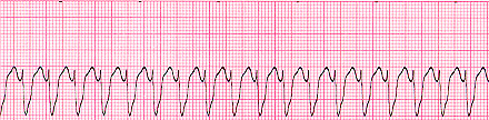

Tmt: Immediate cardioversion-
If stable: Chemical (Amiodarone , Lidocaine) or Sync DC cardioversion
If unstable: has pulse- Synchronised DC cardioversion
no pulse - defibrillation
Note:
Torsades de pointes :
is a different type of VT. The morphology of QRS varies from one complex to the other thus it's refered to as polymorphic VT.
Caused by anything that can prolong QT interval. culprits include Quinidine, Procainamide, Sotalol, Amiodarone (least likely) etc

Tmt includes Magnesium sulphate infusion first!
Tachcardia + wide QRS (diff from a specific SVT called wide QRS SVT, however better to consider and treat as VT until proven otherwise)
Causes include electrolyte abnormalities, MI, drugs, long QT syndrome etc

Tmt: Immediate cardioversion-
If stable: Chemical (Amiodarone , Lidocaine) or Sync DC cardioversion
If unstable: has pulse- Synchronised DC cardioversion
no pulse - defibrillation
Note:
Torsades de pointes :
is a different type of VT. The morphology of QRS varies from one complex to the other thus it's refered to as polymorphic VT.
Caused by anything that can prolong QT interval. culprits include Quinidine, Procainamide, Sotalol, Amiodarone (least likely) etc

Tmt includes Magnesium sulphate infusion first!
Paroxysmal supraventricular tachycardia
Sudden onset palpitation
Dizziness
Chest pain
Dyspnea
+/- LOC
Due to

In WPW, there's the Delta wave- slurred upstroke of the QRS with a short PR interval bcos the signal evades the normal AV node delay and rather passes thru the accessory pathway.

Treatment:
Initial: vagal maneouvres (vasalva, carotid massage)
Drug Tmt :
Adenosine to block AV node except for Wolff-Parkinson-White (AV blockers r contraindicated in WPW becos it'll only promote passage thru accessory pathway.) Amiodarone for WPW
2nd line drugs include verapamil, diltiazem, metoprolol, digoxin
Cardioversion for unstable patients or those not responsive to medications.
Dizziness
Chest pain
Dyspnea
+/- LOC
Due to
- AV node re-entry or
- AV re-entry (av node + accessory pathway)- WPW syndrome

In WPW, there's the Delta wave- slurred upstroke of the QRS with a short PR interval bcos the signal evades the normal AV node delay and rather passes thru the accessory pathway.

Treatment:
Initial: vagal maneouvres (vasalva, carotid massage)
Drug Tmt :
Adenosine to block AV node except for Wolff-Parkinson-White (AV blockers r contraindicated in WPW becos it'll only promote passage thru accessory pathway.) Amiodarone for WPW
2nd line drugs include verapamil, diltiazem, metoprolol, digoxin
Cardioversion for unstable patients or those not responsive to medications.
Conn Syndrome
Primary Hyperaldosteronism
Hypertension
Hypokalaemia
Muscle weakness
Metabolic alkalosis
Hypernatremia
No edema
Adrenal tumour
Diag: Do Aldosterone/ Renin ratio (>30)
Tmt: Spirinolactone (can cause gynecomastia)
Eplerenone
Surgery
Hypertension
Hypokalaemia
Muscle weakness
Metabolic alkalosis
Hypernatremia
No edema
Adrenal tumour
Diag: Do Aldosterone/ Renin ratio (>30)
Tmt: Spirinolactone (can cause gynecomastia)
Eplerenone
Surgery
Sunday, April 29, 2012
Glucagonoma
Presents like Diabete mellitus with Polyuria, Polydypsia, weight loss, hyperglycaemia but with a necrotizing dermatitis- necrolytic migratory erythema(blistering and swelling in areas subjected to friction).
Malignant Pancreatic tumour (alpha cells)
Tmt :surgery
Malignant Pancreatic tumour (alpha cells)
Tmt :surgery
Cystic fibrosis
Defective Chloride transport due to defective CFTR gene (usually a 3base pair deletion)
Aut recessive
Thick mucus secretions causing recurrent sinopulmonary infections, bronchiectasis, FTT
Psedomonas causes Pneumonia (Treat with 2 agents with antipseudomonad coverage)
Pancreatic insufficiency leading to Fat malabsorption with deficiency of vitamins A D E and K, Chronic diarrhoea
Meconium ileus (failure to pass meconium, bilous vomitting, history of polyhydramnios, ground glass appearance on X ray, intestinal perforation)
Positive sweat chloride test (done twice) is diagnostic.
Tmt: high calorie diet, pancreatic enzymes replacement, fat soluble vitamins supplementation. Only life saving tmt is bilateral lung transplant.
Median survival is 30yrs
Pyloric stenosis
Congenital
Infant 4-8 wks old with projectile vomitting which has become more frequent and more forceful
Vomittus is non bilous, occurs after feeding
Visible peristalsis in upper abdo
Failure to thrive
Olive shaped upper abdo mass palpable after vomitting (may however be absent)
Abdo USS confirms diagnosis: shows thick hypoechogenic ring in pyloric area
Tmt: surgery
Intussusception
2yr old Child with abdo pain
presents with features of small bowel obstruction
red currant jelly stool
sausage shaped abdo mass
USS shows target sign (one tube inside another)
Air contast enema both diagnoses and reduces it in most cases
presents with features of small bowel obstruction
red currant jelly stool
sausage shaped abdo mass
USS shows target sign (one tube inside another)
Air contast enema both diagnoses and reduces it in most cases
Paediatric Hip joint pain
Slipped capital femoral epiphysis : Obese adolescent male, referred pain to knee usually present, loss of abduction and internal rotation of hip. Tmt: surgical pinning promptly to avoid avascular necrosis.
Legg-Calve-Perthes disease: child 4-10yrs, male, avascular necrosis of femoral head, idiopathic. Tmt- splint or surgery
Septic arthritis: Emergency. Acute onset, warm swollen jt, hematogenous spread following URTI, staph & strep,
hip is externally rotated, X ray usually normal, wbc count is high, ESR is high.
Do USS guided aspiration- synovial fluid leucocyte >100,000 is definitely SA, do culture.
Tmt : drain immediately, empirical antibiotics nafcillin (or vancomycin ) + cephalosporin
Avascular necrosis in sickle cell anemia: African descent, history suggestive of SS
Transient synovitis: male child 3-10yrs, following trauma or viral infection.
Exclude septic arthritis. if at least 3 of the following are present, do further workup.
Fever>39°C,
WBC >12,000,
ESR >40 ,
Refusal to bear weight
Kawasaki Disease
Vasculitis following infection
Child less than 5yrs
Hyperaemic buccal mucosa, strawberry tongue, Fissured lips
Diagnosis is clinical
Criteria: High fever (>39°C) for 5 days + 4 of ffing: unilateral large lymph nodes,
rash,
mouth changes,
limb desquamation/edema
conjuctivitis
Tmt: IVIG and Aspirin. (unlike other rashes in children where aspirin is contraindicated cos of risk of Reye's syndrome )
May be complicated by Coronary artery aneurysm with resultant risk of MI.
Young child with fever, red eyes, red tongue, red palms and rash
Guillain-Barré syndrome
Ascending paralysis
albumino-cytologic dissociation in CSF(high protein, normal wbc)
Recent Campylobacter jejuni or other infection e.g respiratory
Investigations: nerve conduction studies, LP
Risk of death from respiratory failure
Monitor pulmonary function: Peak inspiratory pressure and FVC
Tmt: IVIG or Plasmapheresis
Subscribe to:
Posts (Atom)